
Family: Bacilladnaviridae
Family: Bacilladnaviridae
Arvind Varsani, Yuji Tomaru, Kenta Okamoto, Anna Munke and Mart Krupovic
The citation for this ICTV Report chapter is the summary to be published as Varsani et al., (2026):
ICTV Virus Taxonomy Profile: Bacilladnaviridae 2026, Journal of General Virology, (in press)
Corresponding author: Arvind Varsani ([email protected]), Mart Krupovic ([email protected])
Edited by: Sead Sabanadzovic and Evelien Adriaenssens
Posted: July 2026
Summary
Bacilladnaviridae is a family of viruses with single-stranded, circular DNA genomes and non-enveloped icosahedral capsids. The genome ranges from 3.4 to 6 kb and contains a short double-stranded region of 0.5–1 kb. All bacilladnavirids that have been cultivated thus far infect diatoms, a group of unicellular algae (Stramenopiles), and cause lysis of the host cells. Uncultivated members of the family have been identified in marine/estuarine organisms and environments. Bacilladnavirid genomes have at least three major open reading frames (ORFs), including ORFs encoding a replication-associated protein and a capsid protein, and appear to replicate by a rolling-circle mechanism. The capsid protein of bacilladnavirids has the jelly-roll fold and is structurally most closely related to the capsid proteins of RNA viruses of the family Nodaviridae. Unlike other viruses in the phylum Cressdnaviricota which have small T=1 capsids, virions of bacilladnavirids have a T=3 icosahedral symmetry.
Table 1 Bacilladnaviridae. Characteristics of members of the family Bacilladnaviridae
| Characteristic | Description |
| Example | Chaetoceros salsugineum DNA virus 1 (AB193315), species Protobacilladnavirus chasesal |
| Virion | Non-enveloped, icosahedral T=3 symmetry, 35 nm diameter |
| Genome | ssDNA(+/-) of 3.4–6.0 kb, 1 circular segment (+) and complementary linear segment(s) (-), thus partially double-stranded |
| Replication | Nuclear, rolling-circle mechanism, lytic |
| Translation | mRNAs produced via DNA transcription |
| Host range | Protists (diatoms) |
| Taxonomy | Realm Floreoviria, kingdom Shotokuvirae, phylum Cressdnaviricota, class Arfiviricetes, order Baphyvirales: 7 genera, 22 species |
Virion
Morphology
Bacilladnavirids form non-enveloped, isometric, icosahedral capsids. Cryo-electron microscopy analysis of Chaetoceros tenuissimus DNA virus type II (CtenDNAV-II, species Protobacilladnavirus tenuis) and Chaetoceros lorenzianus DNA virus (ClorDNAV, species Protobacilladnavirus chaelor) capsids, determined at 2.4 and 2.2 Å resolution, respectively has shown that they have T=3 symmetry (Munke et al., 2022, Gebhard et al., 2025) (Figure 1A Bacilladnaviridae). The capsid protein (CP) subunits adopt a jelly-roll fold. AlphaFold CP structure predictions suggest that the overall capsid architecture is conserved for other bacilladnavirids, although more subtle variation in capsid organization may exist (Varsani et al., 2025). CtenDNAV-II capsid, but not that of ClorDNAV, appears to be stabilized by ions (Munke et al., 2022, Gebhard et al., 2025).
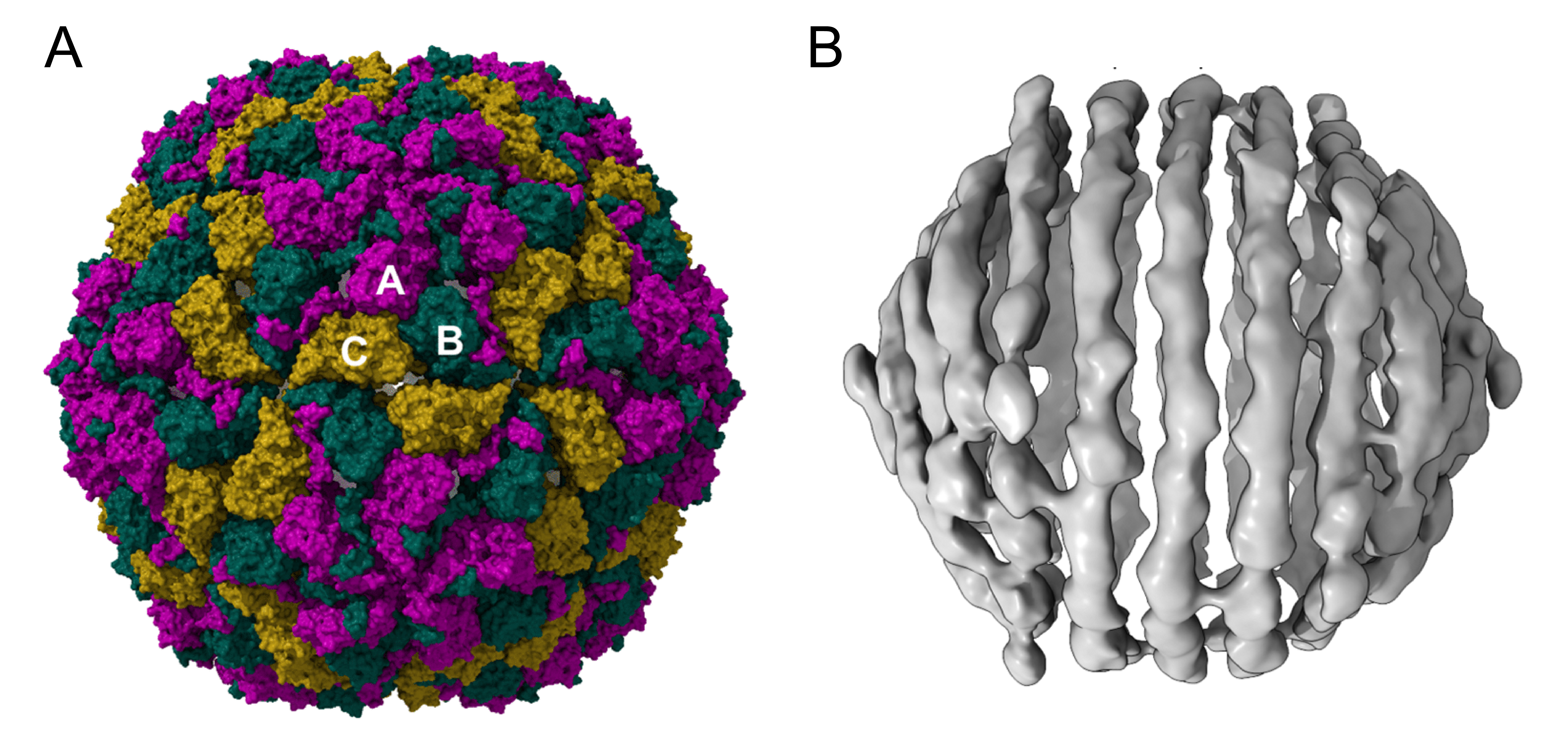 |
| Figure 1 Bacilladnaviridae. (A) Atomic model of the CtenDNAV-II capsid. The three subunits A, B, and C are coloured purple, green, and yellow, respectively. The capsid is rendered with a surface representation viewed down an icosahedral 2-fold axis. (B) The outer genome layer of CtenDNAV-II. The coil of three turns is visualized in the centre of the image, where the individual parallel turns are separated by approximately 28 Å (Munke et al., 2022). |
Physicochemical and physical properties
Virions are relatively stable in the dark at room temperature (Nagasaki et al., 2005, Tomaru et al., 2013b).
Nucleic acid
Bacilladnavirids have a circular single-stranded DNA genome of 3.4–6.0 kb with a shorter, complementary linear segment of 0.5–1.0 kb that creates a double-stranded DNA region (Nagasaki et al., 2005, Tomaru et al., 2011, Tomaru et al., 2012, Toyoda et al., 2012, Tomaru et al., 2013a). However, some potential members of the family may lack the complementary segment, as in the case of the unclassified Chaetoceros debilis DNA virus (Tomaru et al., 2008). The genome is designated as ssDNA(+/-) because the ORFs are encoded in both orientations.
Within the mature virus capsid, the outer layer of the genome appears to adopt a partially spooled arrangement, i.e., tightly wound into concentric layers (Figure 1B Bacilladnaviridae) (Munke et al., 2022, Gebhard et al., 2025). It has been suggested that the observed spool could correspond to the double-stranded region of the genome (Munke et al., 2022). The GC content of the bacilladnavirid genome DNA is 40–50% (Varsani et al., 2025).
Proteins
SDS-PAGE analysis of bacilladnavirid particles has revealed one or two major protein bands (Nagasaki et al., 2005, Tomaru et al., 2008, Tomaru et al., 2011, Toyoda et al., 2012, Kimura and Tomaru 2013, Tomaru et al., 2013b), but the identity of these proteins has not been determined. Structural studies show that the capsid shell is built from a single CP. Comparison of the CPs encoded by viruses with the largest and smallest genomes, Chaetoceros salsugineum DNA virus 1 (CsalDNAV) and Avonheates virus SG_479 (AvVSG_479) shows that the former is 59 aa longer than the latter, with an extended N-terminal region that is enriched in positively-charged amino acid residues oriented into the capsid lumen (Varsani et al., 2025). This “R-arm” may be implicated in genome compaction. The lack of the R-arm in the CPs of the puahadnaviruses AvVSG_479 and Avonheates virus SG_19 (AvVSG_19) suggests a relaxed necessity for the compaction of their smaller genomes. The CP of bacilladnavirids is structurally most closely related to the CPs of positive-sense RNA viruses of the family Nodaviridae (Kazlauskas et al., 2017, Munke et al., 2022).
The virions of many bacilladnavirids have two major structural proteins (Kimura and Tomaru 2015). The second virion protein may be a virus-encoded divergent homolog of phospholipase A1 which could be present in the capsid lumen and involved in virion entry into or egress from host cells at the end of the replication cycle (Varsani et al., 2025).
Genome organization and replication
Bacilladnavirid genomes have 3–5 ORFs. One of these ORFs encodes a replication associated protein (Rep) and a second ORF encodes the CP (Figure 2 Bacilladnaviridae). The remaining ORFs encode proteins of unknown function, although there is evidence that a subset of viruses from the genera Protobacilladnavirus, Diatodnavirus and Aberdnavirus encode a highly divergent homolog of phospholipase A1 (PLA1) (Varsani et al., 2025). Similar to other members of the phylum Cressdnaviricota, the Rep of bacilladnavirids contains two domains: the N-terminal HUH superfamily endonuclease harbouring the three characteristic RCR motifs I, II and III, and the C-terminal superfamily 3 helicase domain containing motifs Walker A, Walker B, motif C and the Arg finger (Kazlauskas et al., 2017). These conserved motifs display signatures that distinguish bacilladnavirids from other members of the phylum Cressdnaviricota. Bacilladnavirid genomes have a conserved gene order (synteny) with the Rep and CP genes in opposite orientations (Varsani et al., 2025).
Based on the presence of the characteristic Rep, bacilladnavirid genomes are believed to replicate by the rolling-circle mechanism initiated by the viral Rep and involving one or more host DNA polymerases. Transcriptomic analysis of CtenDNAV-II-infected cells shows that at the later stages of infection (3 to 24 hours post infection) several components of the host replisome are significantly upregulated. These include DNA polymerase alpha complex (POLA1, POLA2, DNA primase small subunit PRIM1, and large subunit PRIM2), DNA polymerases delta (POLD) and epsilon (POLE), three subunits of ribonuclease H2, and DNA ligase (Hongo and Tomaru 2025). Similarly upregulated are host histones, suggesting that chromatinization of the viral DNA is important for replication.
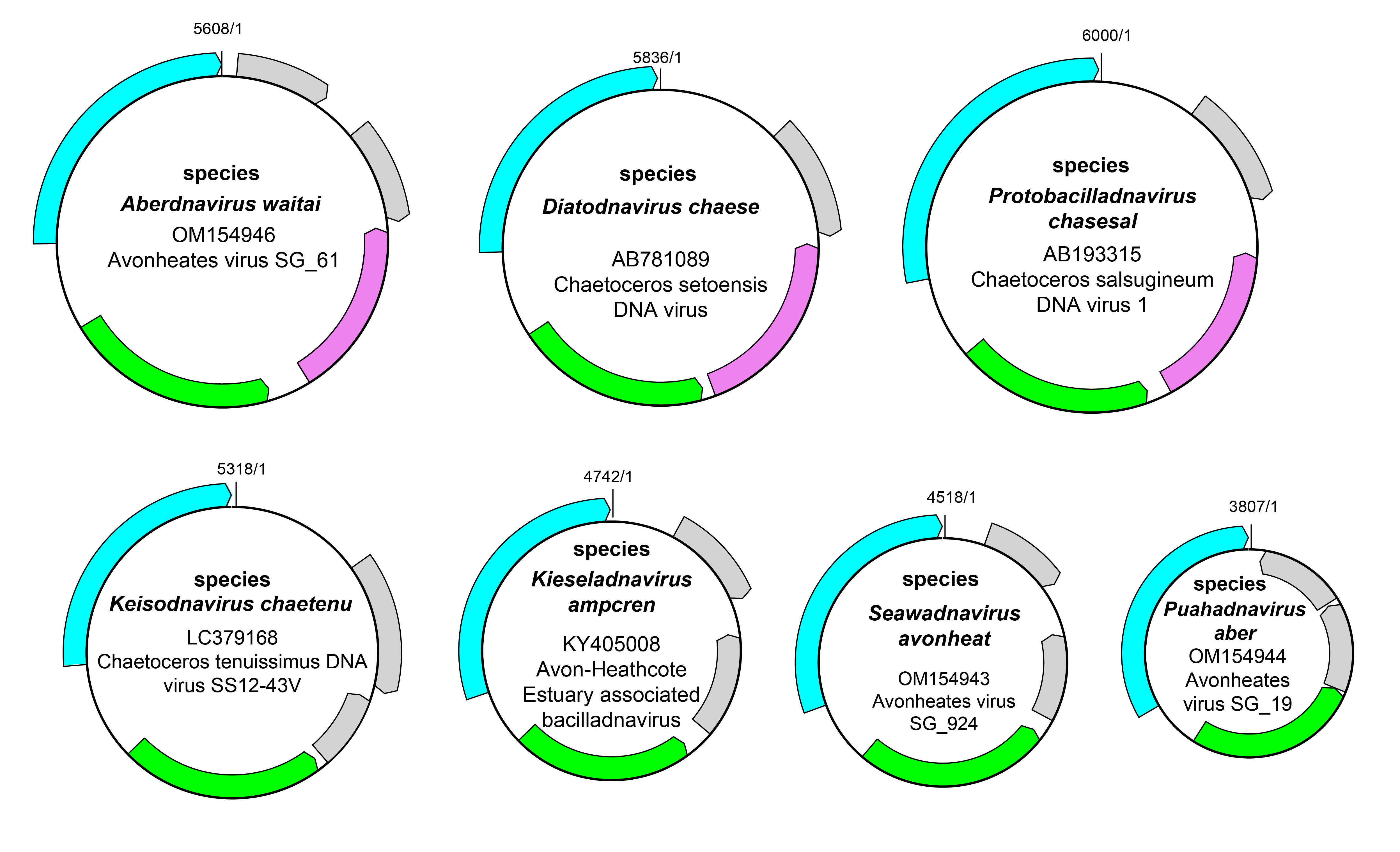 |
| Figure 2 Bacilladnaviridae. Genome organisation of selected viruses representative of the 7 genera in the family Bacilladnaviridae. ORF colours are: Rep, cyan; CP, green, phospholipase, pink, hypothetical proteins, grey. |
Biology
All isolated bacilladnavirids infect diatoms, a cosmopolitan and environmentally important group of unicellular algae (Stramenopiles). Recognized hosts include species from the genera Chaetoceros and Haslea. Bacilladnavirids have also been detected in environmental samples such as ocean water and benthic sediments from estuaries. They have also been identified in samples from invertebrates (mud-flat snails, Amphibola crenata) and vertebrates (red snapper, Lutjanus sp.), but it is most likely that these findings are due to indirect sampling of diatoms (Varsani et al., 2025). Bacilladnavirids have a high host specificity. For example, early studies showed that CsalDNAV was able to infect only one of 58 strains of marine phytoplankton used in experiments (Nagasaki et al., 2005).
Diatoms have a worldwide distribution and an important role in global biogeochemical cycles. The impact of bacilladnavirid infection on the regulation of diatom populations has not been studied in any detail. Viral infection imparts fatal damage on the host in axenic culture but, in nature, diatom host populations maintain their sizes despite the presence of the infectious viruses. This may imply diverse virus resistance strategies but these have not yet been discovered (Tomaru et al., 2015).
Derivation of names
Aberdnavirus: from the Welsh aber, meaning estuary and DNA for deoxyribonucleic acid
Bacilladnaviridae: from Bacillariophytes, the host name for many viruses in this family and DNA for deoxyribonucleic acid; the suffix -viridae for family taxa
Diatodnavirus: from diatom-infecting ssDNA virus
Keisodnavirus: from the Japanese Keisō (珪藻), meaning diatom and DNA for deoxyribonucleic acid
Kieseladnavirus: from the German Kieselalge, meaning diatom and DNA for deoxyribonucleic acid
Protobacilladnavirus: from the Ancient Greek πρωτο- (prōto-) meaning first, and ssDNA virus infecting Bacillariophytes
Puahadnavirus: from the Māori pūahatanga, meaning estuary and DNA for deoxyribonucleic acid
Seawadnavirus: from seawater and DNA for deoxyribonucleic acid
Bacilladnavirid species epithets are derived from host species or isolation sources using names in local languages (Varsani et al., 2025).
Demarcation criteria for taxa within the family
The classification of genera within the family Bacilladnaviridae is based upon the phylogenetic analysis of the Rep protein (Kazlauskas et al., 2017) with genera representing distinct monophyletic clades.
Relationships within the family
The family Bacilldnaviridae includes 7 genera; Aberdnavirus, Diatodnavirus, Keisodnavirus, Kieseladnavirus, Protobacilladnavirus, Puahadnavirus and Seawadnavirus based upon a phylogenetic comparison of the Rep protein (Figure 3 Bacilladnaviridae).
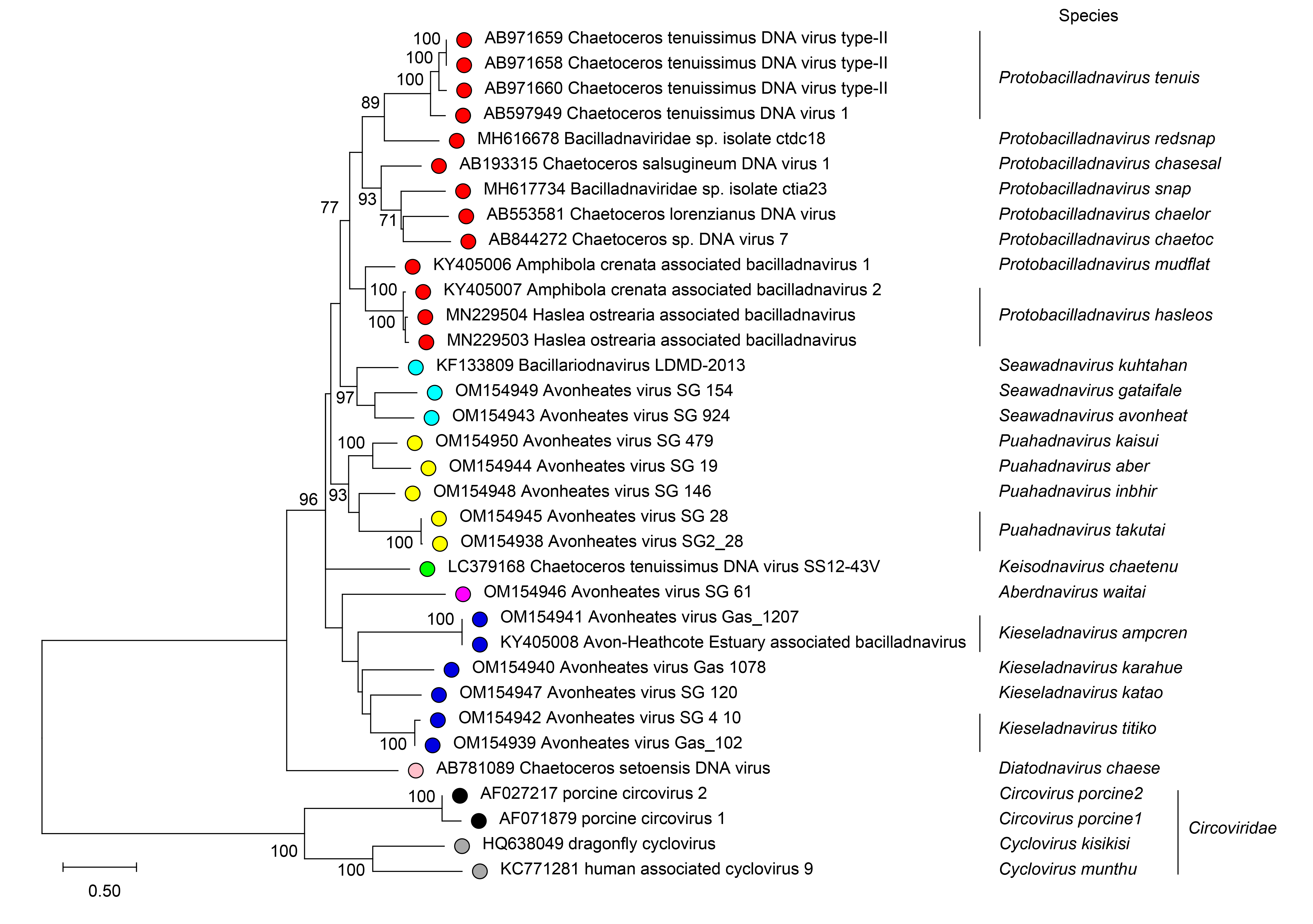 |
| Figure 3. Bacilladnaviridae. Evolutionary history of the of bacilladnavirid Rep proteins. Rep sequences aligned using MAFFT (Katoh and Standley 2013) were used to infer maximum likelihood phylogenetic trees using PhyML 3.0 (Guindon et al., 2010) with best-fit model rtREV+G+I determined using ProtTest 3 (Darriba et al., 2011). Representative circovirus sequences were used to root the tree. Numbers at branches indicate bootstrap support where this was >70%. Scale bar: number of substitutions per site. |
It should be noted that although the genera defined by analysis of the Rep protein are monophyletic, analysis of the CP protein produces a different phylogeny, which can be interpreted as a consequence of intergeneric recombination within the family (Varsani et al., 2025).
Relationships with other taxa
Bacilladnavirids comprise a phylogenetically distinct clade of ssDNA viruses. The conserved motifs of bacilladnavirid Rep proteins display signatures that distinguish bacilladnavirids from other members of the phylum Cressdnaviricota (Kazlauskas et al., 2017, Krupovic et al., 2020); namely, the FP and PF residues in the motif I and Arg finger, respectively, as well as the two glutamates in the Walker B motif, instead of the aspartates present in the Reps of most other cressdnaviricots. Furthermore, bacilladnavirids are distinguished from most other known cressdnaviricots in that they form T=3 capsids, which are considerably larger that T=1 capsids of other members of the phylum.
Related, unclassified viruses
| Virus name | Accession number | Virus abbreviation |
| bacilladnavirus sp. isolate ctcc592 | MH617605 | BacVctcc592 |
| Chaetoceros tenuissimus DNA virus_GoT | OR283035 | CtenDNAV_GoT |
| Chaetoceros debilis DNA virus 83 | LC379166 | CdebDNAV83 |
Virus names and virus abbreviations are not official ICTV designations.